- #How to identify exon and intron in sequence bioedit update
- #How to identify exon and intron in sequence bioedit full
This review aims to highlight the importance of studying variation in deep intronic sequence as a cause of monogenic disorders as well as hereditary cancer syndromes.Īndrogen Insensitivity Syndrome Duchenne Muscular Dystrophy Fabry Disease Splice Enhancer Splice Site. Additionally, deep intronic mutations can disrupt transcription regulatory motifs and non-coding RNA genes.

Deleterious DNA variants located more than 100 base pairs away from exon-intron junctions most commonly lead to pseudo-exon inclusion due to activation of non-canonical splice sites or changes in splicing regulatory elements. Here we review evidence from mRNA analysis and entire genomic sequencing indicating that pathogenic mutations can occur deep within the introns of over 75 disease-associated genes. Yet, for a substantial proportion of patients, sequence information restricted to exons and exon-intron boundaries fails to identify the genetic cause of the disease.
 Each exon/intron will have a live link to the corresponding genomic sequence.įor more information, see this video tutorial.Next-generation sequencing has revolutionized clinical diagnostic testing. If you are working with eukaryotic genes that have transcript variants, select the variant that you want and click on + to expand the exon table.
 #How to identify exon and intron in sequence bioedit full
Change the record display from Full Report to Gene Table (the display menu is located on the left side above the record). Use the Tools button to display Sequence Text View. Zoom on the part of the sequence of your interest (for example an exon of eukaryotic gene, indicated as a solid box in the graphics). falciparum intron residues are A or T ( 11 ). NCBI Sequence Viewer (graphics) displays the gene region as a line. The canonical 5GU-AG 3 sequence was almost 100 conserved, and the immediately flanking exon sequences had a consensus sequence of WR for the 5 and RW for the 3 splice site (Table 2). Select the genomic record that you want, and choose between the GenBank or FASTA Download. To learn more about the NCBI RefSeqGene project, please see the project's home page and this factsheet. To locate the RefSeqGene sequence, check for the NG_ genomic sequence. If you are working with a human gene, check if one of the available genomic sequence is named the RefSeqGene sequence. There will be more than one Genomic section if there is an alternative genomic locus representing the gene. Scroll to the section(s) labeled Genomic. If the independently determined RefSeq mRNA cannot be aligned perfectly to the genome, the lengths given in the Gene Table display may differ from that of the mRNA sequence itself. The link will take you to the section of the Gene record that reports RefSeqs for the gene. The report of intron/exon organization in the Gene Table display is based on the placement of exons and CDS on the genomic sequence. Select the link to the NCBI Reference Sequences (RefSeq) from the Table of contents. By expanding the region, you will be able to obtain upstream/downstream sequence of the gene. #How to identify exon and intron in sequence bioedit update
If you want to adjust the range to capture, modify the values in the Change region shown tool on the FASTA display and click on Update View.
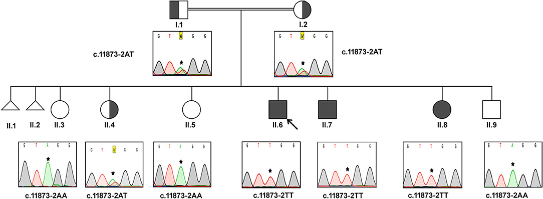
The FASTA format will display the sequence. To learn more about the sequence display formats, please see the following factsheet. The GenBank display -not to be confused with a GenBank record- will display a flat file with annotation, followed by the sequence in numbered rows. Cloned genomic DNA is prepared for inverse polymerase chain reaction (PCR) by digesting the DNA with a restriction enzyme and circularizing the. This unit describes identifying intron/exon boundaries in genomic DNA by comparing nucleotide sequences of genomic DNA to cDNA. Select the record display format that you want. Identification of intron/exon boundaries in genomic DNA by inverse PCR. A heterozygous genomic 7-base deletion in intron 2 of the SOD1 gene was identified in two KC families.  Use Go to nucleotide: Graphics FASTA GenBank.
If there is an alternative genomic locus representing the gene, the Genomic Sequence information will expand into a pull-down menu, allowing you to select an alternative genomic record.Intron regions were sampled more extensively because we hypothesized, before conducting the experiment, that these regions are most polymorphic. Scroll to the Genomic regions, transcripts, and products section. exon and intron or noncoding 5regions were identified in the se-quence.Select the Full Report display ( example).(If you are retrieving too many records, see these tips for searching for genes.) There are a few methods that will allow you to obtain the genomic sequence for the gene: Find the gene record that you want in the NCBI Gene database.


 0 kommentar(er)
